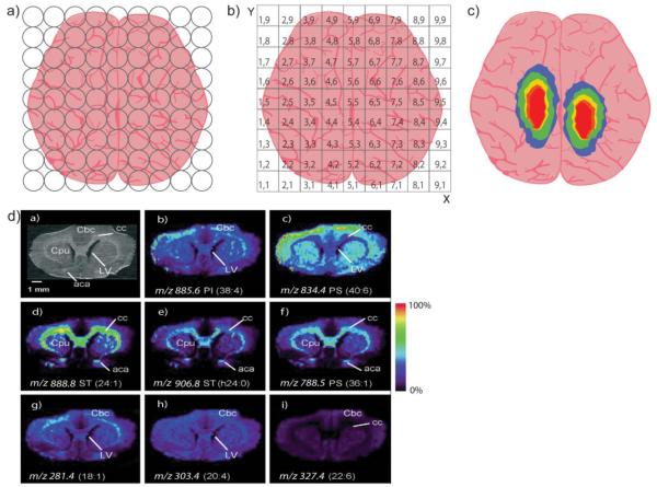
Scheme 1. Creating and Imaging Ions by Mass Spectrometry.
(a) After tissue is prepared, mass spectra are collected at points across the entire tissue. (b) Each sampled point is converted into spatial coordinates and (c) an image is created by displaying the intensity of a specific ion at each point. (d) Real images of 8 different lipids acquired simultaneously from a rat brain, reprinted with permission from [11].

