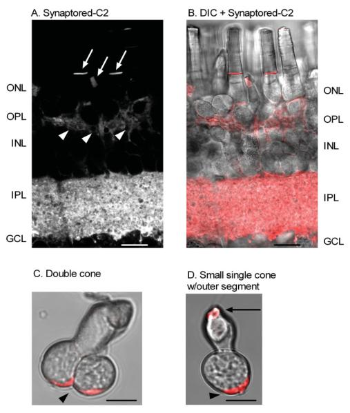
Fig. 3.
Activity-dependent dye (Synaptored-C2) loading in photoreceptor terminals of the tiger salamander retina. Synaptored-C2 activity-dependent labeling is restricted to terminal regions in the OPL and IPL of the retinal slice preparation. A: A living retinal slice loaded with 40 μM Synaptored-C2 for 15 min in the dark, then washed with ADVASEP-7 (500 μM). Arrowheads indicate synaptic loading of Synaptored-C2 into rod and cone terminals. Arrows indicate Synaptored-C2 dye uptake into outer segments of rods and cones. B: Combined brightfield differential interference contrast (DIC) image and Synaptored-C2 labeling of the retinal slice in A. C: Brightfield image of an isolated double cone loaded with Synaptored-C2 and then enzymatically dissociated with papain. D: Brightfield image of an isolated small single cone with outer segment loaded with Synaptored-C2 and then enzymatically dissociated with papain. In isolated cells, arrowheads indicate synaptic uptake of the activity-dependent dye, Synaptored-C2, and arrow indicates dye uptake into outer segment as seen in A and B of the retinal slice. Scale bars = 10 μm.

