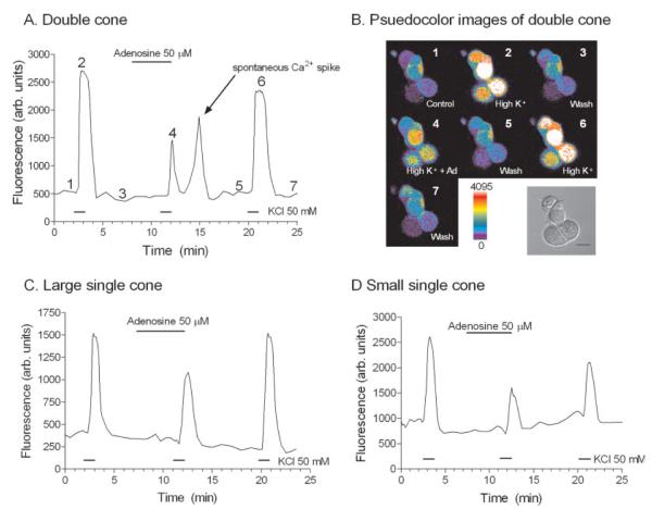
Fig. 5.
Adenosine inhibits the K+-evoked-depolarization in terminals of isolated cone photoreceptors. A: A plot of [Ca2+]i changes in the terminal of a double cone. B: Corresponding pseudocolor images from the double cone plotted in A, with a brightfield DIC image of the isolated double cone. Fluo-4-loaded double cone displayed in B: 1, control, normal (2.5 mM) amphibian superfusate; 2, elevated (50 mM) [K+]o containing amphibian superfusate; 3, wash or normal amphibian superfusate; 4, adenosine (50 μM) and elevated [K+]o containing superfusate; 5, wash, normal amphibian superfusate; 6, elevated [K+]o containing amphibian superfusate; and 7, wash, normal amphibian superfusate. The numbers correspond to time points of image acquisition of A in B. All measurements were made from regions of interest drawn over the terminal and defined as the broad synaptic vesicle-filled extension located under the soma in the perikaryal region (see Materials and Methods). All cells were stimulated with a 1-min application of elevated (50 mM) [K+]o. C: [Ca2+]i measurements from a large single cone terminal. D: [Ca2+]i measurements from a small single cone terminal. Scale bar = 10 μm.

