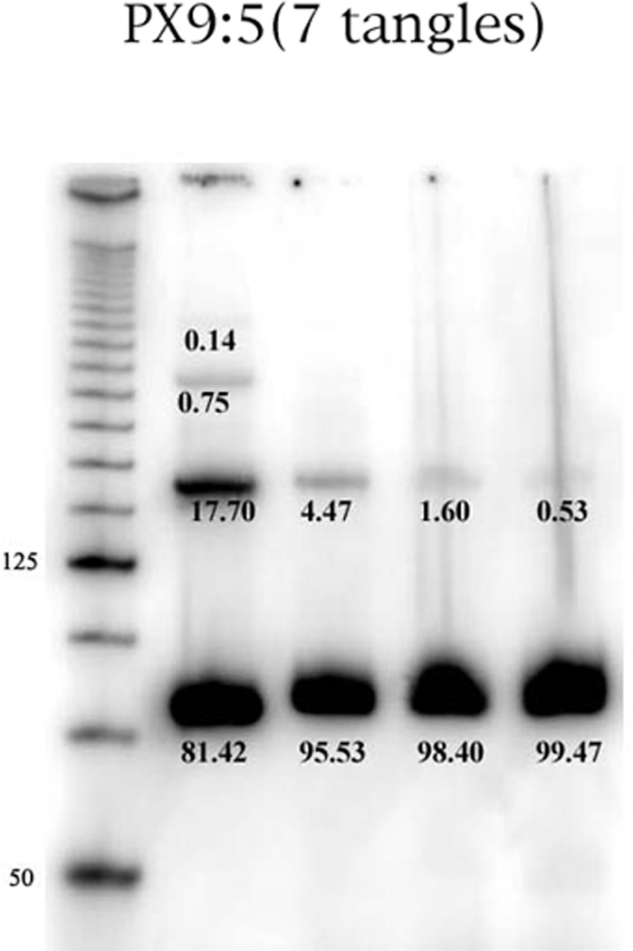
Figure 7. Concentration Dependence of the 9:5 PX Motif.
This is an autoradiogram of a PX 9:5 complex containing 7 half-turns of DNA. The leftmost lane contains a 25 base-pair marker. From left to right, the other lanes contain the complex at concentrations of 1000 nM, 500 nM, 250 nM and 125 nM. The percentage of material in each band is indicated. It is clear that the multimers of the 9:5 PX virtually can be eliminated at low concentrations.

