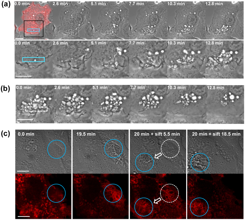
Figure 1. Photo-manipulation of PA-Rac1 reversibly controls membrane ruffling and macropinosome formation.
(a) Time-lapse imaging of RAW264 cells expressing mCherry-PA-Rac1 during photoactivation using 445 nm laser irradiation. RAW264 cells were transiently transfected with pTriEx/mCherry-PA-Rac1 and subjected to local photoactivation of PA-Rac1 (blue rectangular area). The first panel shows a merged image of phase-contrast and mCherry fluorescence before the photoactivation of PA-Rac1, confirming the expression of mCherry-PA-Rac1. The elapsed times after starting the irradiation are shown at the top of each panel. The lower panels show magnified images of the boxed area of the cell. The bar indicates 10 μm. (b) Time-lapse series of the same region of the cell shown in Fig. 1a after ceasing irradiation. Elapsed times after ceasing the irradiation are shown at the top of each panel. The bar indicates 10 μm. (c) DIC (upper) and mCherry (lower) images of the mCherry-PA-Rac1 expressing cell irradiated at two different positions sequentially. Time 0 indicates the initiation of blue laser irradiation in the area enclosed by a blue circle (the first panel). At 19.5 min, ruffles were apparent within the irradiated region (blue circle in the second panel). After 20 min, the irradiation was shifted to a different area of the same cell. After ceasing the irradiation, the ruffles immediately receded, and spherical macropinosomes were formed in the initial irradiation area (broken-lined circle in the third panel). Instead, marked ruffling was induced in the new irradiated area (blue circles in the third and fourth panels). The bar indicates 10 μm.

