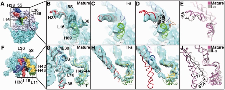
Figure 4.
Conformational variability of H38 of the 23S rRNA in the 45S intermediates. Structural comparison of H38 in the I-a (B–E) or II-a (G–J) structures, with that of the mature 50S structure. Two different thumbnails of the mature 50S subunit (A and F), with a few components highlighted in different colors, are shown in left panels. (B, C, G and H) Close-up view of the mature 50S (B and G), the I-a (C) and II-a (H) structures in the same area as in their respective thumbnails, with their atomic models superimposed. (D and I) The I-a (D) and II-a (I) structures are superimposed with the atomic model of the mature 50S structure. (E and J) Conformational change of H38 between the mature and the I-a (E) or II-a (J) state. The 5S rRNA, H38, H89 and H42–44 are colored blue, red, green and yellow, respectively. Ribosomal proteins, L16, L36, L30 and L11 are colored black, golden, purple and dark green, respectively. For clarification, the 23S rRNA helices colored gray in the upper left thumbnail (A) are not shown in (B–E).

