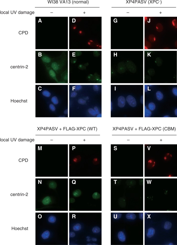
Figure 3.
Subcellular localization of endogenously-expressed centrin-2. Indicated cell lines were subjected to double immunofluorescent staining with anti-CPD (red) and anti-centrin-2 (green) antibodies with or without localized UVC irradiation. The nuclear DNA was counter-stained with Hoechst 33342.

