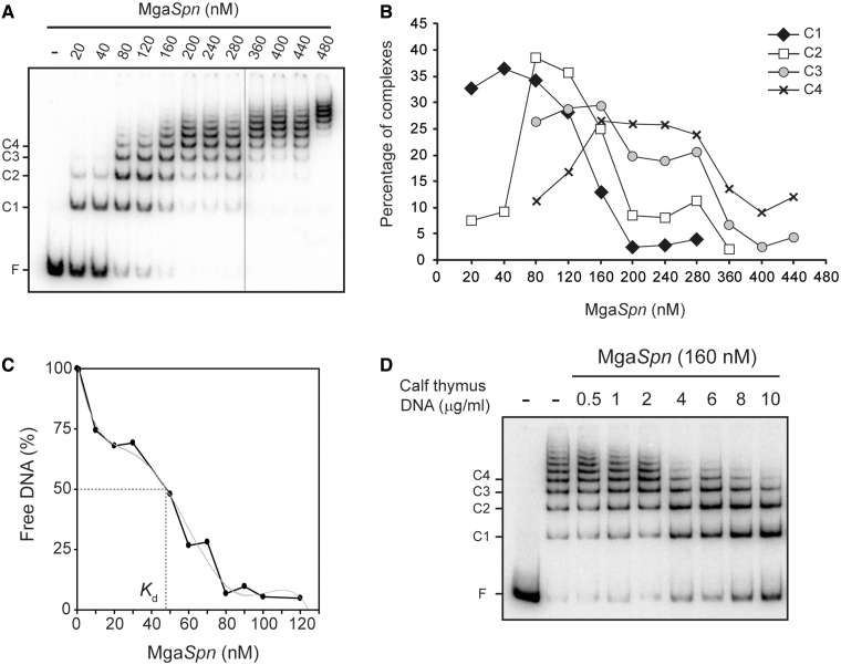
Figure 3.
Formation of multimeric MgaSpn-DNA complexes. (A) EMSA of MgaSpn-DNA complexes. The 32P-labelled 222-bp DNA fragment (2 nM) was incubated with increasing concentrations of MgaSpn in the presence of non-labelled competitor calf thymus DNA (2 µg/ml). Reactions were loaded onto a native gel (5% polyacrylamide). All the lanes displayed came from the same gel. Bands corresponding to free DNA (F) and to several MgaSpn–DNA complexes (C1, C2, C3, C4) are indicated. (B) The autoradiograph shown in A was scanned, and the percentage of the indicated complexes was plotted against the concentration of MgaSpn. (C) Affinity of MgaSpn for the 222-bp DNA. The labelled 222-bp DNA (0.1 nM) was incubated with different concentrations of MgaSpn (1–130 nM) in the absence of competitor DNA. Free DNA and bound DNA were separated by native gel electrophoresis. The percentage of free DNA was plotted against MgaSpn concentration. (D) Dissociation of higher-order MgaSpn-DNA complexes. The indicated amount of non-labelled calf thymus DNA was added to pre-formed MgaSpn-DNA complexes (160 nM MgaSpn, 2 nM labelled 222-bp DNA).

