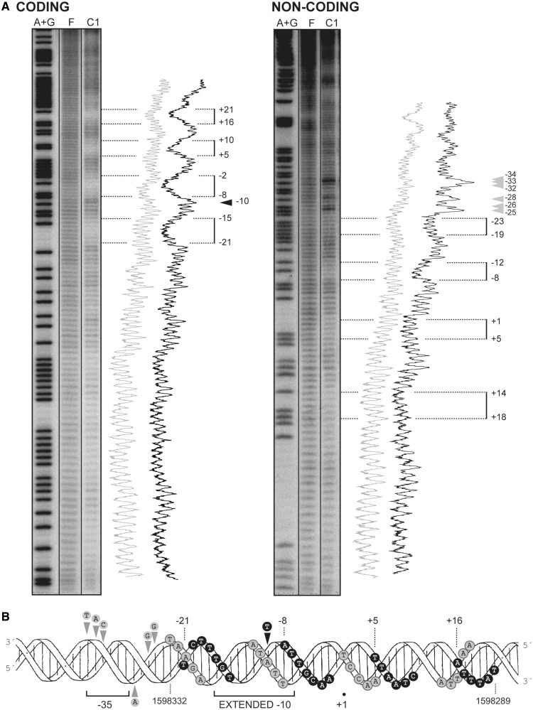
Figure 7.
MgaSpn binds preferentially to the Pmga promoter region on the 224-bp DNA. (A) Hydroxyl radical cleavage pattern of the 224-bp DNA without protein (F) and with MgaSpn bound to its primary site (complex C1). Coding and non-coding strands relative to the Pmga promoter were 32P-labelled at the 5′end. Lanes A + G are products from Maxam-Gilbert adenine- and guanine-specific sequencing reactions performed on the respective labelled strands. All the lanes displayed came from the same gel. Lane C1 corresponds to a longer exposure time. Densitometer scans from lanes F (grey line) and C1 (black line) are shown. Numbers indicate positions relative to the transcription start site of the Pmga promoter. Regions protected by MgaSpn are indicated with brackets. Arrowheads indicate positions more sensitive to hydroxyl radical cleavage. (B) B-form DNA of the Pmga promoter region showing MgaSpn contacts as deduced from hydroxyl radical. Black and grey circles indicate protein contacts on the coding and non-coding strand, respectively.

