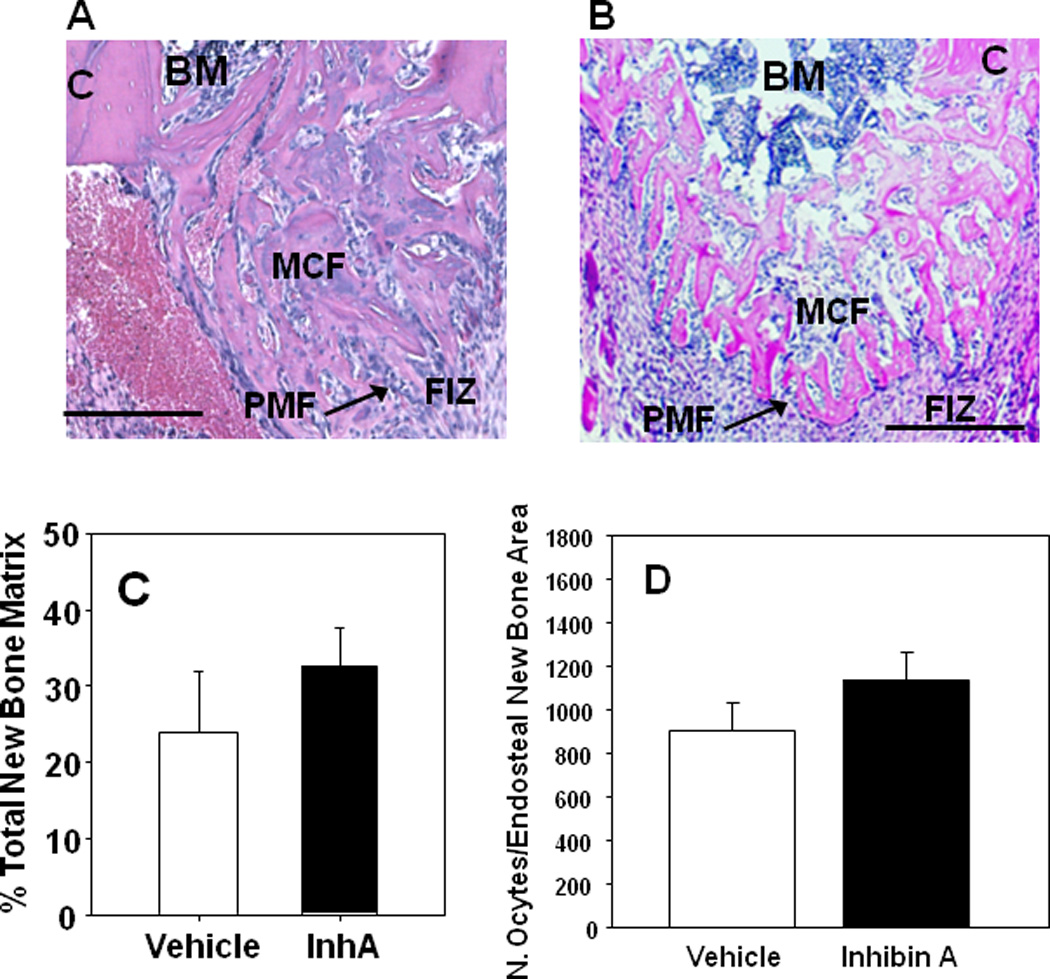
Figure 3. hInhA overexpression enhanced new microcolumn formation but not the area of new matrix formation during DO.

Photomicrographs of the proximal half of a decalcified paraffin embedded histological sections of the DO gap in Glvp/InhA mice treated with Vehicle during DO (A), or overexpressing hInhA during the time of DO (B) and stained with H&E depicting the major biological zones of the distraction gap. Analytically, the zone of microcolumn formation (MCF) is referred to as Endosteal New Bone (ENB). BM, Bone Marrow; C, Cortex; FIZ, Fibrous Interzone; PMF, Primary Matrix Front. Bars = 500µm. (C) The formation of new bone matrix in the endosteal distraction gap of Vehicle-treated Glvp/InhA mice and Glvp/InhA mice treated with MFP was assessed in central sections of decalcified DO gaps stained with H&E. (D) The osteocyte density in the ENB was enumerated per bone area in central sections of decalcified DO gaps stained with H&E. No significant differences in cellularity were observed. Data shown are mean +/− SD.
