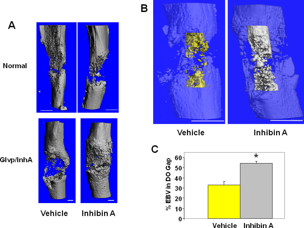
Figure 4. hInhA overexpression increases endosteal new bone formation during DO.

(A) MicroCT reconstruction of mouse tibiae. (Top) Swiss-Webster treated with vehicle or hInhA for 18 days. (Bottom) Glvp/InhA mice treated with Vehicle or MFP to overexpress hInhA. Mice were sacrificed 18 days post-operatively and the DO gap scanned. (B) MicroCT translucent reconstructions illustrate hInhA effects on endosteal bone volume. 3-Dimensional reconstruction of endosteal new bone in Vehicle treated (yellow) and InhA overexpressing (gray) Glvp/InhA mice. (C) Total volume and bone volume of the endosteal distraction gap were calculated directly from the voxel volumes in the reconstruction. Data shown are mean +/− SD; *=P<0.05.
