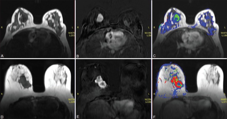
Figure 2 (A-F).
A 36-year-old female with lump in right breast. Axial non–fat-suppressed pre-contrast VIBE image (A) and subtracted VIBE image (B) reveal an enhancing mass in the right breast. (C) Color overlay images for the Ktrans value of the right breast lesion reveal low values suggestive of a benign lesion. HPE: Fibroadenoma. Axial non–fat-suppressed pre-contrast VIBE image (D) and subtracted VIBE image (E) reveal an irregular enhancing mass in the right breast. (F) Color overlay images for the Ktrans value of the right breast lesion reveal high values suggestive of a malignant lesion. HPE: IDC

