
Figure 18 (A-C).
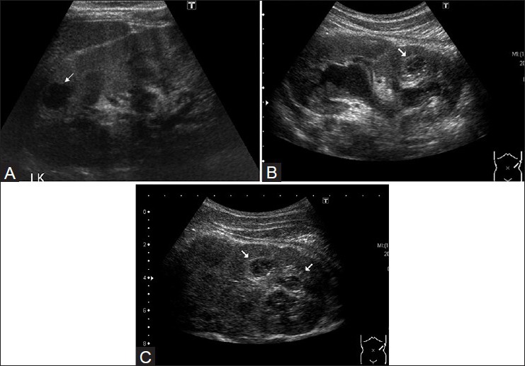
(A) High-resolution ultrasound images (acquired with a 7.5 MHz transducer) demonstrate a small irregular caseous cavity (white arrow) in the upper part of the left renal parenchyma, (B) high-resolution ultrasound images revealing a tuberculous cavity with fine septae within, in the lower part of the left kidney of another patient. Note marked urothelial thickening in this dilated system, (C) USG image revealing irregular sonolucent cavities, with a semisolid echo texture