
Figure 23 (A-C).
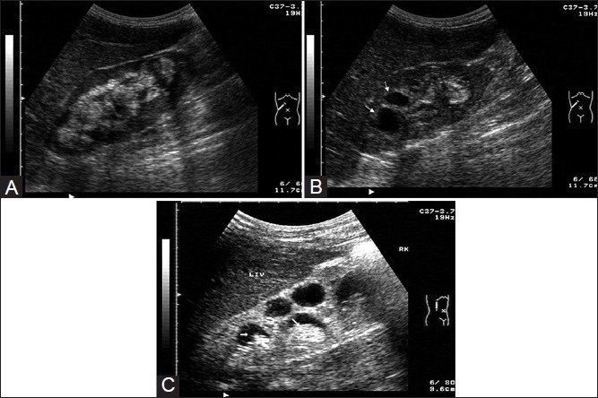
(A) USG image revealing hyperechogenic areas of caseation interspersed with the echogenic sinus echoes. (coronal scan), (B) Oblique USG scan reveals uneven caliectasis (white arrows) with a hazy interface and urothelial thickening in the upper calyces. The lower calyceal region is replaced by hyperechogenic caseous tissue, (C) Comparative USG image of regular (evenly dilated) caliectasis with hyperechoic fungal balls (white arrows) in a HIV-positive patient (note the hyperechogenic material is lying within clearly dilated calyces and are not replacing them as happens in tuberculous caseation)