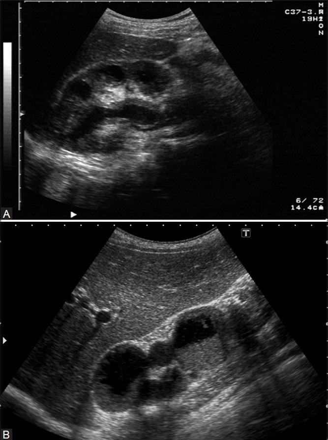
Figure 24.

(A) Moderate-to-severe urothelial thickening noted throughout the visualized urothelium. This is well visualized on account of the dilatation due to a tuberculous ureteric stricture, (B) USG image revealing uneven caliectasis with ragged urothelial thickening (arrowheads). Note significant debris in the lower calyces
