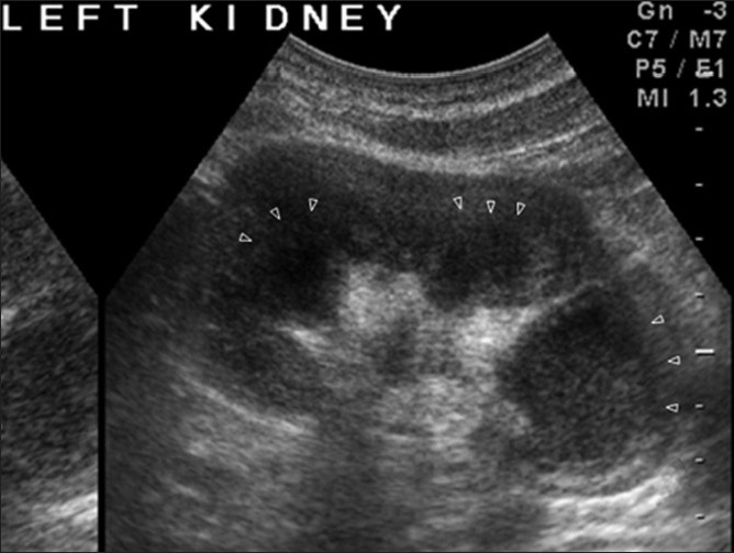
Figure 26.

USG image showing evolution of tuberculous lobar caseation. Different phases of destruction are apparent. (Lower group calyces are completely merged with the parenchyma, midgroup calyces about to merge, and upper ones almost merged). Arrowheads demarcate the junction between residual parenchyma and the dilated calyces
