
Figure 27.
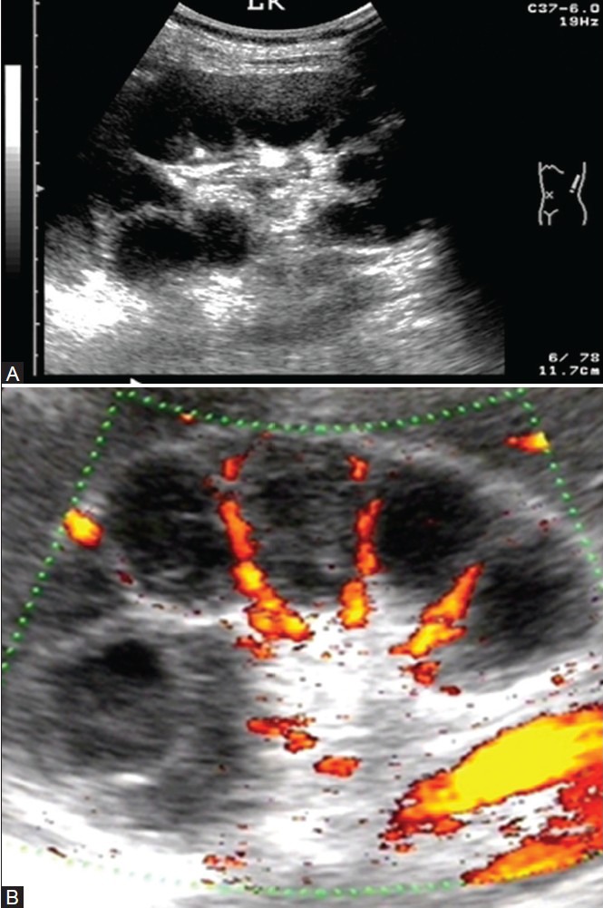
(A) USG image revealing lobar caseation (A) Grey scale and, (B) Color flow image demonstrating presence of renal vasculature only between the caseated lobes
Official websites use .gov
A
.gov website belongs to an official
government organization in the United States.
Secure .gov websites use HTTPS
A lock (
) or https:// means you've safely
connected to the .gov website. Share sensitive
information only on official, secure websites.
(A) USG image revealing lobar caseation (A) Grey scale and, (B) Color flow image demonstrating presence of renal vasculature only between the caseated lobes