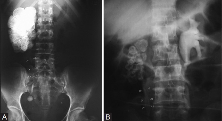
Figure 3 (A, B).

(A) Plain radiograph revealing classic lobar pattern of calcification, which is pathognomonic of end-stage renal tuberculosis. Ureteral calcification is also noted, which is fainter in upper parts (arrowheads), (B) intravenous urogram revealing the ‘classic’ lobar pattern of calcification in a non-functioning (R) kidney. (The lobar distribution of calcification is better appreciated in the upper half of the kidney). Ureteral calcification is also noted (arrowheads)
