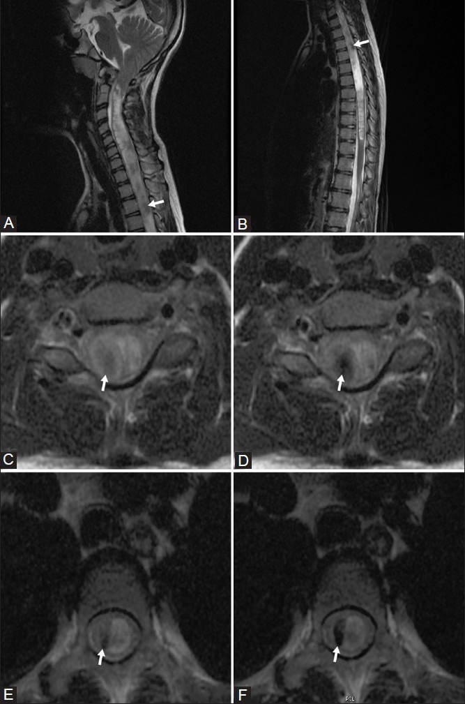
Figure 1 (A-F).

Sagittal T2-weighted images showing syrinx in cervical (A) and thoracic (B) cord; faint flow voids (arrows) are seen within the syrinx. (C, D): Axial T2-weighted images of cervical cord with (c) and without (d) flow compensation; flow voids (arrows) are more obvious in D (without flow compensation). (E, F): Axial T2-weighted images of thoracic cord with (E) and without (f) flow compensation; flow voids (arrows) are more obvious in F (without flow compensation)
