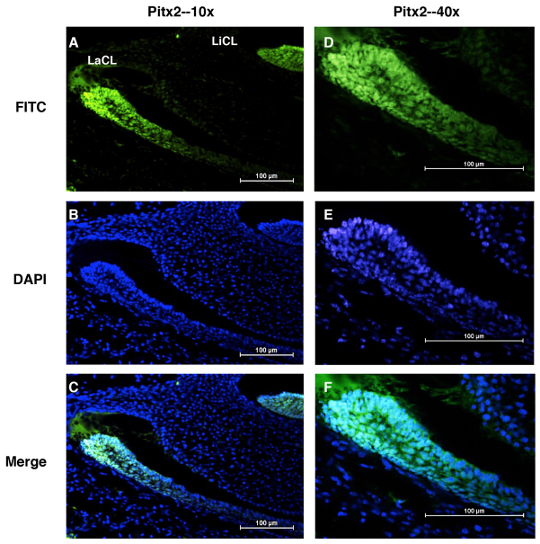
Fig. 5.
Pitx2 expression in the E16.5 mouse incisor. (A-C) Pitx2 staining of embryonic day (E) 16.5 mouse incisor showing increased Pitx2 expression (green) in the labial cervical loop (LaCL) and lingual cervical loop (LiCL), which contain dental stem cells. DAPI staining (blue) was used to visualize the nuclei and provide contrast. (D-F) Higher magnification of A-C. Original magnifications are indicated.

