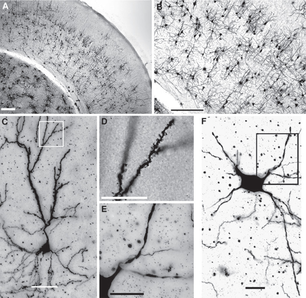
Fig. 1.
Golgi-stained section of the barrel cortex. a Low magnification image (×4) illustrating barrel cortex and its six distinct layers bounded by pia and the white matter. The white box highlights the region magnified in b; Scale bar 250 µm. b Higher magnification (×10) highlighting the morphologies of layer 6 (L6) neurons. Scale bar 250 µm. c Higher magnification (×20) of a single pyramidal neuron located in L6. Scale bar 50 µm. d High magnification (×100) of the region in white in c. Please note the presence of dendritic spines, demonstrating the entirety of the neuronal labeling. Scale bar 50 µm. e High magnification (×100) of the region in black in f. Please note the relative absence of dendritic spines. Scale bar 25 µm. f ×20 magnification of a typical labeled non-pyramidal neuron located in L6. Scale bar 25 µm

