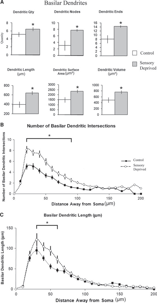
Fig. 5.
Effect of chronic sensory deprivation on basilar dendritic parameters of L6 pyramidal neurons. a Basilar dendritic morphometric variables in control versus sensory-deprived animals. Basilar dendritic parameters showed an overall trend to increase significantly (including dendritic nodes, dendritic ends, and dendritic length) following chronic sensory deprivation in developing animals; means and one standard error of the mean are plotted. b Mean number of basilar dendritic intersections between 10-µm concentric spheres in control versus sensory-deprived mice. The increased number of dendritic intersections in sensory-deprived relative to control mice was distributed from 20 to 50 µm away from the center of the soma, suggesting that the effect is relatively localized. c Mean number of basilar dendritic lengths between 10-µm concentric spheres in control versus sensory-deprived mice. Similar to mean number of basilar dendritic intersections, the decreased basilar dendritic length in sensory-deprived relative to control animals was distributed mostly from 30 to 60 µm away from the soma, indicating the localized effect of sensory deprivation on the development of basilar dendrites. Error bars indicate standard error of the mean (SEM). Asterisks indicate statistical significance of post hoc tests (Fisher LSD) at individual levels (p < 0.05)

