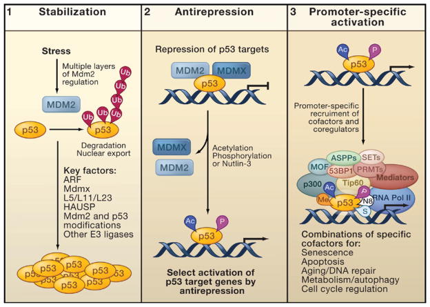
Figure 4. Refined Model for p53 Activation.
Promoter-specific p53 activation in vivo consists of three key steps: (1) p53 stabilization, (2) antirepression, and (3) promoter-specific activation.
(1) Stress-induced p53 stabilization occurs through many different mechanisms, many of which act by affecting the ability of Mdm2 to ubiquitinate p53.
(2) Antirepression describes the release of p53 from the repression mediated by Mdm2 and MdmX. This step requires the acetylation of p53 at key lysine residues and facilitates the activation of specific subsets of p53 targets. Phosphorylation of p53 or treatment with the small molecule Nutlin-3 may have similar effects on antirepression.
(3) For full activation of specific promoters, p53 recruits and interacts with numerous cofactors. These act by modifying p53, the surrounding histones, or other transcription factors. Regulating the activation of specific groups of p53 targets for apoptosis, senescence, cell cycle control, DNA repair, autophagy, metabolism, or aging may require exact combinations of cofactors and posttranslational modifications.
Abbreviations: Ac, acetylation; P, phosphorylation; Me, methylation; N8, neddylation; S, sumoylation.

