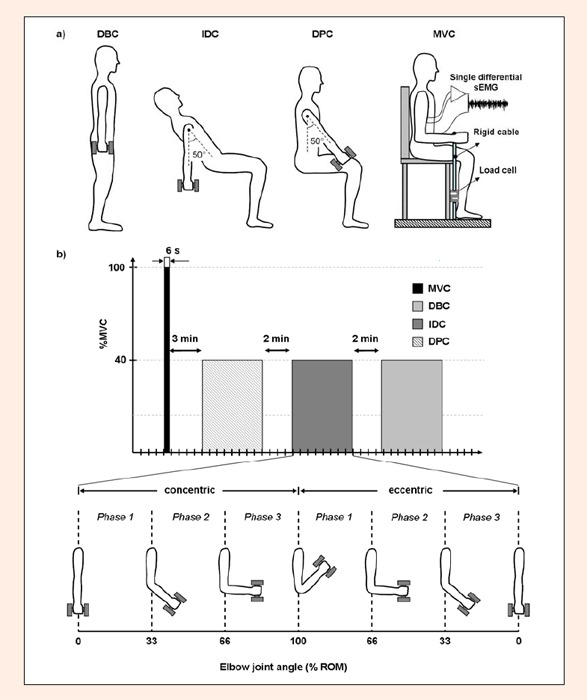
Figure 1.

Schematics of the experimental setup. a) Body orientation for each dumbbell curl protocol and for the MVC trial including electrodes placement; b) Time sequence of each test trial (randomized) and rest periods; c) Partition of the dumbbell curls cycle into concentric and eccentric contractions and further division into three phases according to elbow joint angle.
