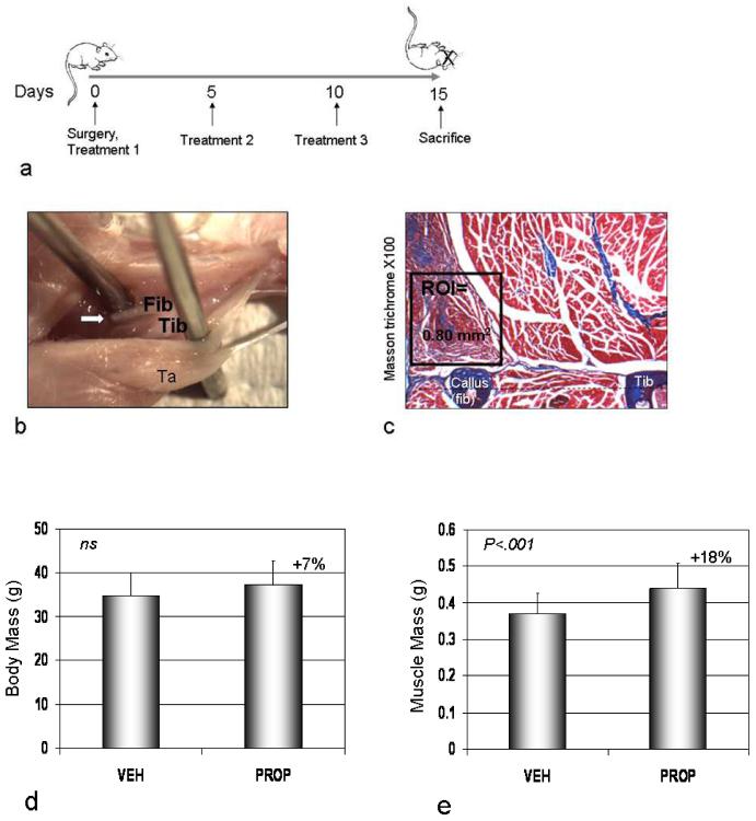
Fig. 1.
(a) Mice received treatments on the day of surgery, 5 days post-op, 10 days post-op, and were euthanized 15 days following the initial treatment. (b) A fibula osteotomy procedure was used (arrow) and the lateral compartment muscles were cut. Fib-fibula, Tib-tibia, TA-tibialis anterior. (c) Histological sections at the osteotomy site were stained using Masson trichrome and 0.80 mm2 region of interest lateral to the fracture callus examined for fraction of fibrotic tissue (blue). (d) Body weight and (e) muscle mass (b; triceps brachii + quadriceps femoris) in saline (VEH) and propeptide (PROP; 20 mg/kg) treated mice. Error bars represent S.D.

