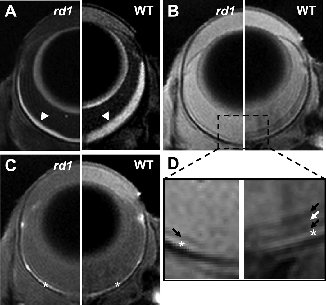
Figure 1.
Diffusion weighted (A), non-diffusion weighted (B), and T1-weighted images of the eyes of an rd1 (left side) and a WT (right side) mice. The zoom-in view of non-diffusion weighted image (D) shows a dark retinal layer in the rd1 mouse eye and three retinal layers exhibiting dark-bright-dark signal intensity in the WT mouse eye. Arrow heads indicate retina/choroid complex; arrows indicate MR-detected retina layers; * indicates the choroid.

