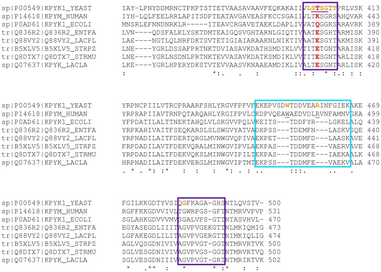
Figure 2. Section of the multiple sequence alignment of PYK showing the C-domain with the allosteric site.
The boxed sequences directly contribute to the allosteric binding site. The residues in the purple boxes contribute to the phosphate binding site referred to here as 1′Pibs and those in the cyan box to 6′Pibs. The residues underlined in purple within the 1′Pibs site form a structural P-loop motif as discussed by Hirsch and colleagues [30]. The residues marked in orange correspond to the residues that interact with the allosteric ligand, FBP, in the Saccharomyces cerevisiae PYK (1A3W). The LAB PYKs show a conserved glutamate residue at the center of the allosteric site highlighted in red. In Saccharomyces cerevisiae PYK, it was shown experimentally that the mutation of T403 to E403 prevents allosteric activation of this PYK.

