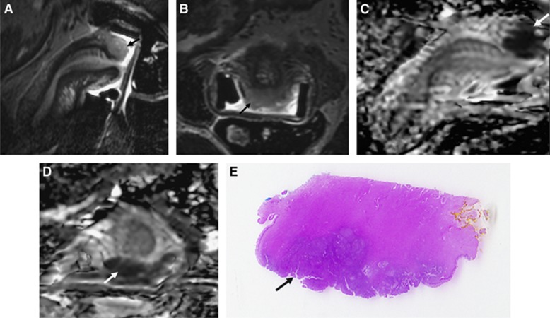
Figure 1.
True positive in a 25-year-old female diagnosed with stage I carcinoma of the cervix following intermittent postcoital bleeding. T2-W images in the sagittal (A) and coronal (B) planes orthogonal to the cervix show a small cervical tumour (arrows). The ADC maps generated from ZOOM-DWI in corresponding orientations (C, sagittal and D, coronal) show that the tumour (arrows) is more easily appreciated in C and D, although considerable image distortion is present. This mandates the use of the T2-W images for anatomical delineation. Good correspondence is seen with the histological definition of the tumour (dark blue) on the haematoxylin and eosin-stained corresponding trachelectomy specimen (E, arrow).

