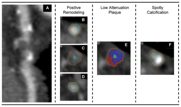
Fig. 1.

Coronary CT images of one patient with a high-risk Lesion. Curved Multi-planar Reconstructed images (A) demonstrated a coronary lesion with >50% luminal narrowing and all three high-risk features. Quantitative analysis assessing outer wall dimensions (orange line in: B, proximal reference; C, at the lesion; D, distal reference) yield a positive remodeling index of greater then 1.05. Volumetric measurements of the entire lesion demonstrated 28.5% low attenuation plaque (attenuation <30 HU, red super imposed in a representative, cross-sectional image [E]). Further, a spotty calcification (<3 mm) was adjunct to the lesion (F). If at least two of the three plaque features were present at the site of a coronary lesion, it was considered as high-risk lesion. (For interpretation of the references to color in this figure legend, the reader is referred to the web version of the article.)
