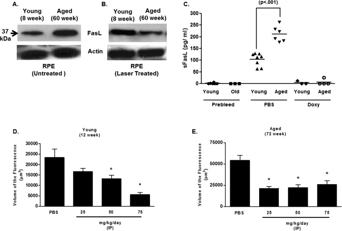
Figure 2.
FasL expression in the eye and blood of young and aged mice. Western blotting for FasL expression in isolated RPE cells from young (8 weeks) and aged (60 weeks) was performed on (A) untreated or (B) laser treated mice. Actin expression was used as the loading control for the Western blots. (C) sFasL levels in serum were determined 48 hours following laser treatment in PBS-treated or doxycycline (Doxy)-treated mice (50 mg/kg/day beginning on the day of laser). Levels were determined using a commercially available ELISA (see Methods). Levels below the detection limit were coded as 0 pg/mL and were included in the analysis. (D) Young (12 weeks) and (E) aged (72 weeks) mice were laser treated and then injected daily by an IP injection of doxycycline at the indicated dosage. The volume of CNV lesions (expressed as volume of the fluorescence) was assessed on day 7. Asterisk (*) denotes significantly different from PBS-treated control.

