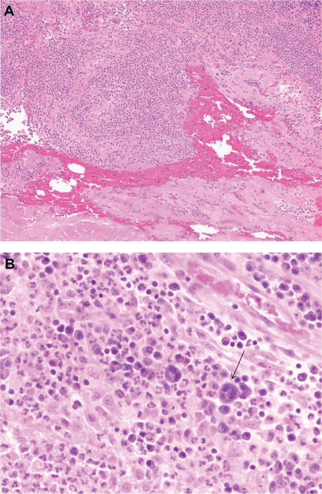
Figure 6.

(A) Histopathology results for a specimen taken from the T2/3 level of the back (lead insertion wound). Microscopic analysis reveals granulation tissue with hemorrhage, acute and chronic inflammation, and a focal foreign body giant cell reaction. (B) Arrow points to multinucleated giant cell seen close up from tissue detailed in (A).
