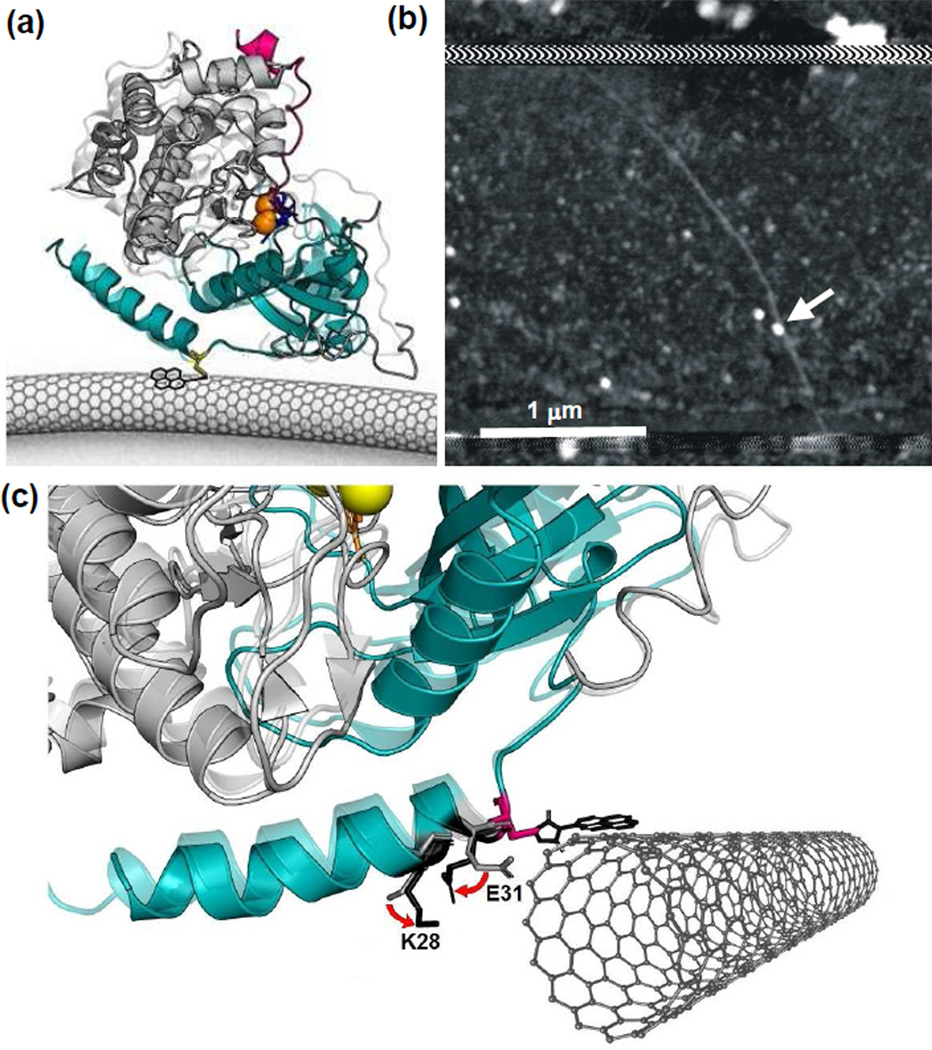
Figure 1.
PKA-labeled, SWNT-based nanocircuits for single molecule electronic monitoring (a) Schematic of a single core catalytic subunit of PKA (small lobe in cyan and large lobe in gray) attached to an SWNT-based circuit through a single cysteine (yellow). Two magnesium ions (orange) position ATP (blue) in the binding pocket. As PKA catalyzes the transfer of γ-phosphate from ATP to its substrate, Kemptide (magenta), the protein undergoes a conformational change from the open (opaque, PDB: 1J3H) to the closed (transparent, PDB: 1ATP) state. (b) Example atomic force micrograph of a SWNT device with a single protein attachment (arrow). Horizontal stripes at top and bottom show the extent of a passivating polymer that covers the distant source and drain electrode connections. (c) Schematic diagram of the PKA-SWNT interface, showing close-up X-ray crystal structures of PKA in its closed (opaque) and open (transparent) conformations (PDB: 1ATP and 1J3H, respectively). The C32 attachment to the pyrene-maleimide linker molecule provides a fixed reference point. In the vicinity of C32, residues 28 and 31 have charged side chains that move appreciably from the open configuration (light gray) to the closed conformation (black). The pyrene and SWNT are independently free to rotate around the C32 site, but this illustration features an energetically likely orientation.

