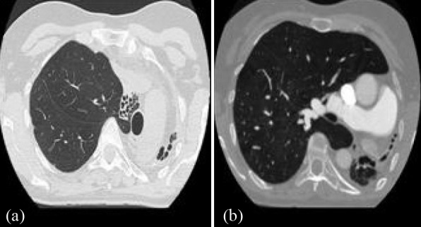
Figure 5. Case 3: Chest CT at the level of the aortic arch (a) demonstrates the hypoplastic left lung associated with the ipsilateral mediastinal shift and multiple bronchiectasis. Chest CT at the level of the pulmonary artery (b) demonstrates the enlarged right main branch.

