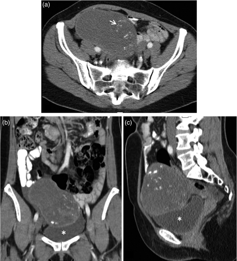
Fig. 2.
On axial contrast-enhanced CT scan (a) was seen a large heterogeneous solid mass, with areas of low attenuation and dystrophic calcifications, suggestive of mucin content. Coronal (b) and sagittal (c) reformatted CT images, revealed the mass located superior to the bladder and extending through the right flank. The lesion contact the bladder but no focal thickening of the bladder wall was evident

