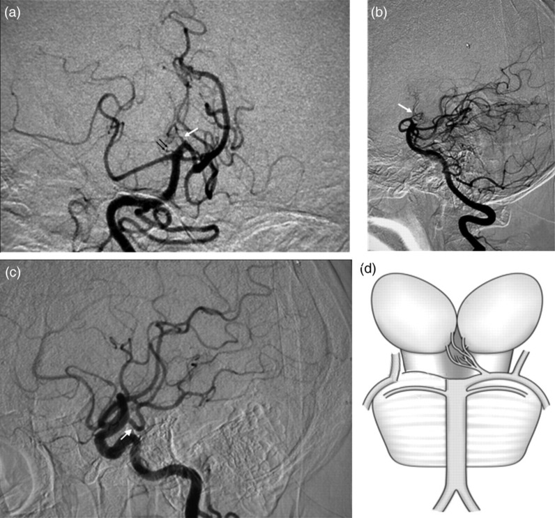
Fig. 2.
(a, b) DSA of the right vertebral injection, anteroposterior view (a) and lateral view (b) showed a stenotic AOP originating from the left P1 segment (white arrow). The P1 segment of the right PCA was absent (black double-arrow). (c) DSA of the right carotid artery injection showed the abnormally enlarged right PcomA could be the source of the right PCA (white arrow). (d) A schematic diagram represents a possible new variant of AOP. The P1 segment of the right PCA is absent while the AOP originates from the left

