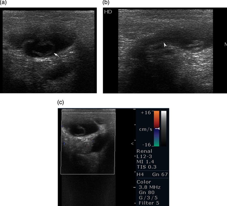
Fig. 1.
Ultrasonography of the right inguinal area was performed with a 7–10 MHz linear array transducer. (a) Transverse view shows well-defined, sausage-shaped, hypoechoic lesion with small internal septations (arrow). (b) Longitudinal view shows sausage-shaped lesion with tail directed cranial and posteriorly (arrow head). (c) Color Doppler showing no internal or peripheral vascularity of the lesion

