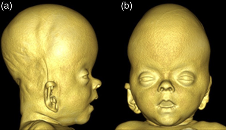
Fig. 4.
3D reconstruction of the craniofacial soft-tissues based on the CT scanning performed at 9 weeks of age. (a) Profile view of the head. Note the extreme head shape with decreased length and increased height. Also note the protrusion of the forehead and the depressed nasal bridge. (b) Frontal view of the head. Note the bulging temporal regions and the hypertelorism

