. 2012 Jan-Mar;16(1):51–56.

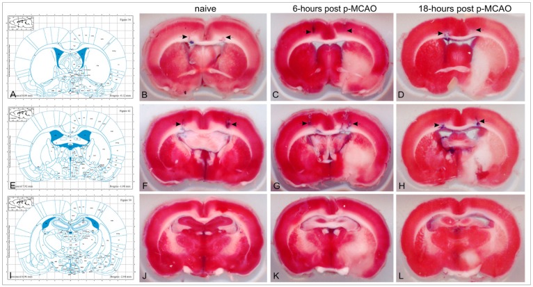
Figure 2. The corresponding Paxinos atlas' plates (with permission from "The rat brain in stereotaxic coordinates", Paxinos G. and Watson C., figures 34, 42 and 50; copyright Elsevier Academic Press 2005) and the representative brain TTC-stained sections at the levels -0.12 (B, C, D), -1.08 (F, G, H) and -2.04mm (J, K, L) from bregma. Blue traces on the TTC sections (arrows) indicate the track and final position of the stereotaxic needle. Naïve animals (group N) did not need any correction of the Paxinos coordinates (B, F). On 6-hours, the classical coordinates (uncorrected) were sufficient enough for invasion in the lateral ventricles of p-MCAO animals (subgroup Av) (C, G). On 18-hours (subgroup Bv), the new (corrected) coordinates were needed to successfully reach the targeted ventricle in p-MCAO animals (D, H).

Figure 2