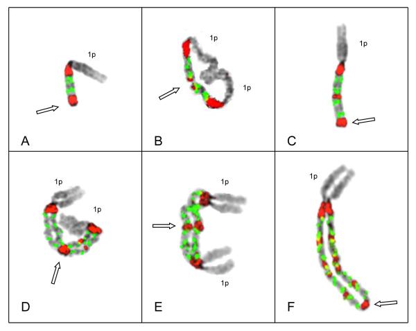
Figure 1.
Representative metaphase chromosomes demonstrating key structures in the progression of intrachromosomal amplification of 1q12~23. Probes for satII/III (red) and CKS1B (green) are shown on inverted DAPI images of chromosomes depicting G-band-like patterns. Patient # 66 demonstrated doublets (two green signals) of CKS1B (see text for explanation) in all cells analyzed. (A) Chromosome 1 in an early stage of amplification, shows four copies of CKS1B bracketed by two copies of satII/III and deletion of chromosome 1q distal to the amplicon. Open arrow demarks the point of sister chromatid fusion (SCF) at distal end. (B) Isodicentric chromosome 1 with two copies of 1p and two copies of the amplicon, both bracketed by satII/III yielding a total of 8 copies of CKS1B. Open arrow indicates the SCF point on the distal 1q (demarked by the satII/III signals) around which 1q hinges in an anaphase bridge. (C) Derivative chromosome 1 showing one copy of 1p and two copies of the amplicon. This chromosome demonstrates the deletion of the 1p distal to the two amplicons (open arrow), and the SCF point for the next step of cycle (open arrow). (D & E) Examples of isodicentric chromosomes 1 from different cells, both showing four amplicons, yielding 16 copies of CKS1B. Note a copy of the short arm of chromosome 1 on each end and the large middle satII/III signals (open arrow) in these chromosomes demarks the point of SCF. (F) Extended ladder-like structure consisting of five copies of the amplicon containing doublets of CKS1B (CN of 18) and one copy of 1p. In this chromosome the lack of sister chromatid cohesion is apparent except at the distinct SCF at the most distal copy of satII/III (open arrow).

