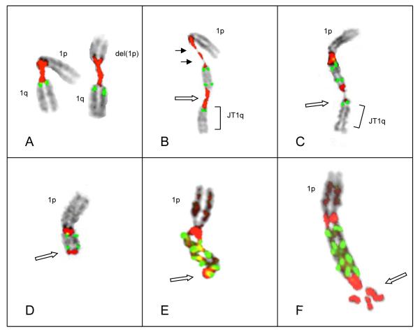
Figure 3.
(A) Patient # 28 demonstrated pericentromeric decondensation in both chromosomes 1, each with a single copy of CKS1B. The chromosome 1 on the left has a normal 1p, while chromosome1 on right has a visible interstitial deletion of 1p. (B) An inverted duplication subsequently occurred in the normal chromosome 1 yielding three copies of CSK1B, associated with two decondensed regions of 1q12 sat II/II. Note the decondensation and near-breakage (solid arrows) in proximal satII/III signal, and decondensation in distal satII/III signal (open arrow). (C) Chromosome 1 with breakage in distal satII/III signal. The satII/III region (open arrow) shows red signal on both sides of thread-like unstained chromatin which appears to break away and result in either a JT1q (bracket) or possibly a micronucleus. (D) Chromosome 1 showing inverted duplication, with two copies of CKS1B bracketed by satII/III, and the loss of an entire 1q distal to the inversion (open arrow), as in Patient #64 (Fig 2C) (E&F) Examples of chromosomes 1 showing ladder-like structures each showing two amplicons bracketed by satII/III signals. Note deletion of 1q distal to the amplicons (open arrows).

