Figure 4.
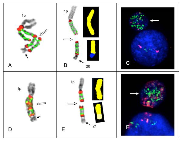
Secondary aberrations including unbalanced translocations and/or the formation of micronuclei subsequently results in the masking of amplicon ladders. Translocation of intact copies of amplicons to non-homologous chromosomes and loss of amplicons by micronucleus formation was found in both Patients # 66 and 64. (A) In Patient #66 an amplified chromosome 1 demonstrating a ladder of four amplicons yielding 16 copies of CKS1B showing a small non-homologous chromosome (solid arrow) fused at the most distal satII/III signal. Breakage is occurring in the satII/III signals in one chromatid (open arrow) midway between the centromeres of the two chromosomes. This unstable dicentric is the putative precursor to chromosomes shown in (B). (B) An unbalanced translocation of two amplicons in patient # 66 is characterized by both FISH (left) and SKY (right) in the same cell. FISH hybridization (on left) shows two copies of the amplicon remaining on donor chromosome 1, while a section of 1q also containing two copies of the amplicon is translocated to a section of receptor chromosome 20 (on right) identified by SKY. Breakage in the middle of the amplicon ladder between the centromeres of this unstable dicentric ladder resulted in eight copies of CKS1B remaining on the der(1), while 8 copies were translocated to der(20). (C) Chromosome 1 from Patient # 64 showing chromosome 21 fused to distal satII/III signal with two copies of the amplicon (CKS1B CN 4). Open arrow indicates the putative breakage point between the centromeres of this dicentric which would result in the chromosomes shown in (D). FISH signals (left) show one amplicon copy remains on the donor chromosome 1 and one copy of the amplicon is translocated to the receptor chromosome 21. SKY (right) confirms one copy of the amplicon is translocated to the chromosome 21. (E and F) DAPI counterstaining demonstrates micronuclei (arrows) from Patients # 66 (E) and # 64 (F) showing high CNs of CKS1B in micronuclei, presumably resulting from the loss of acentric copies of amplicons (not shown).

