
Figure 5. Analysis of peripheral vessel branching patterns in the mouse OIR model.
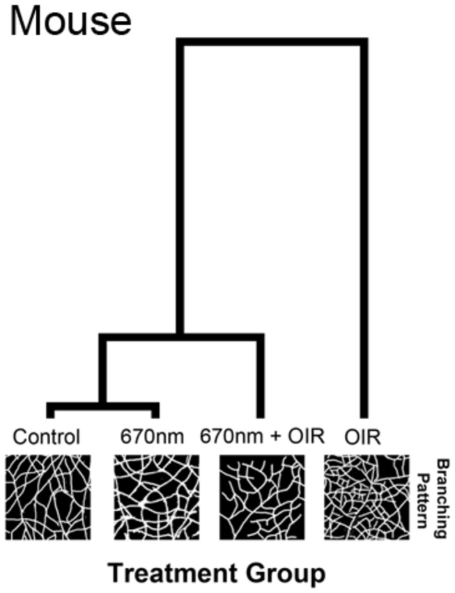
The hierarchical clustering diagram indicates a divergence from the experimental groups with the largest divergence occurring with the OIR group. The Mahalanobis distance was calculated from a MANOVA procedure and is shown relative to control. The OIR group clusters alone (1.96 distance) indicate a very different pattern of peripheral vasculature, to the other experimental groups. 670nm+OIR (0.46 distance) reduced the alteration in the peripheral patterning to almost that of control and 670nm (0.32 distance). The length of the lines in the hierarchical clustering diagram indicate the relative difference between groups.