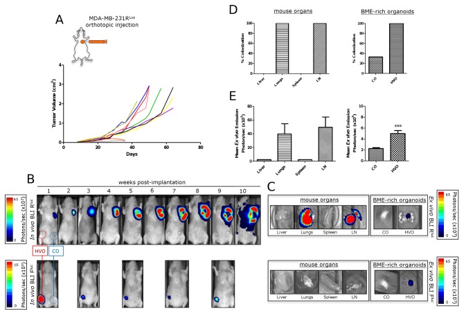
Figure 5. Spontaneous metastasis from primary mammary fat pad tumors.
MDA-MB-231RLuc human breast cancer cells were implanted into the orthotopic environment of the left axillary MFP of nude mice bearing human vascularized BME-rich organoids (HVO) and control BME-rich organoids (CO), without human cells. Primary tumor growth (a, b), metastatic spread (b-e) and HVO functionality (b, c) were monitored over time in vivo and ex vivo. (a) Tumor growth curves for MDA-MB-231RLuc (n = 10) orthotopic mammary tumors. (b) In vivo ventral coelenterazine-based RLuc-BLI images (upper panels) and D-luciferin-based FLuc-BLI images (lower panels) of a representative mouse. (c) Ex vivo coelenterazine-based RLuc-BLI images (upper panels) and D-luciferin-based FLuc-BLI images (lower panels) of excised lungs, inguinal lymph node (LN), spleen, liver, HVO and CO of a representative mouse. (d) Percentage of metastatic colonization and (e) tumor burden in harvested tissues (normal mouse organs and BME-rich organoids) of mice (n = 8) with primary tumor growth. Significant differences (*** p < 0.001).

