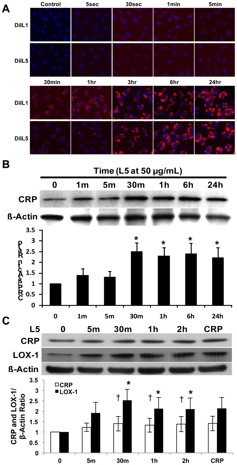
Figure 5. Time-course analysis of L1 and L5 internalization by human aortic endothelial cells (HAECs) and L5-induced C-reactive protein (CRP) expression.
(A) Results of fluorescence microscopy showing that DiI-L1 and DiI-L5 (each red) were internalized by HAECs at different time points. The nuclei of HAECs were co-stained with 4′,6-diamidino-2-phenylindole (DAPI) (blue). (B) Western blot showing that L5 augmented endothelial CRP expression in a time-dependent manner. Time point 0 represents the phosphate-buffered saline (PBS) control. *P<0.05 vs. PBS-treated control. (C) L5 deprivation study showed that internalized L5 continued to induce CRP and lectin-like oxidized receptor-1 (LOX-1) expression 24 hours after replacing L5 conditioned culture media with fresh EGM2 media. L5 exposure times are shown. HAECs were incubated with recombinant CRP for 2 hours as a positive control. Time point 0 represents the PBS control. For all experiments, n = 3, and bars in graphs represent standard deviation. †P<0.05 vs. PBS-treated CRP control, *P<0.05 vs. PBS-treated LOX-1 control.

