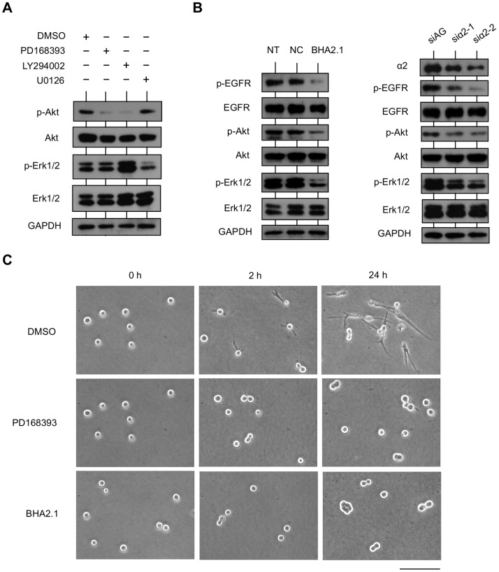
Figure 6. EGFR and integrin α2β1 coordinately regulate downstream signaling pathways responsible for IR cell invasion.
(A) Regulation of EGFR in PI3K/Akt and MEK/Erk1/2 signaling pathways. IR cells in 3D collagen gel were treated with DMSO (control), EGFR inhibitor PD168393 (10 µM), PI3K inhibitor LY294002 (50 µM), or MEK inhibitor U0126 (10 µM) for 24 h. Cells were harvested, and the amounts of phosphorylated and total Akt (Ser473) and Erk1/2 (Tyr202/Tyr204) were analyzed by western blotting. (B) Regulation of integrin α2β1 on EGFR, PI3K/Akt, and MEK/Erk1/2 signaling pathways. Left: IR cells in 3D collagen gel were non-treated (NT), treated with non-function blocking antibody for integrin α2β1 (NC), or treated with function blocking antibody for integrin α2β1 (BHA2.1) for 24 h. Right: IR cells that transfected with siRNA specific to AzamiGreen (siAG) as a negative control or a siRNA specific to integrin α2 (siα2-1 or siα2-2) were transferred to 3D collagen gel and cultured for 24 h. Cells were harvested, and the amounts of integrin α2, phosphorylated and total EGFR (Tyr1068), Akt (Ser473), and Erk1/2 (Tyr202/Tyr204) were analyzed by western blotting. (C) Inhibition of EGFR or function blocking of integrin α2β1 in cells invading collagen. IR cell suspension in medium with DMSO, PD168393, or BHA2.1 was seeded on collagen gel. Phase contrast images for each sample at 0 h, 2 h, and 24 h are shown. DMSO-treated IR cells have already protruded into collagen at 2 h and present long protrusions at 24 h. In contrast, PD168393- or BHA2.1-treated IR cells simply adhere to the collagen with a spherical morphology.

