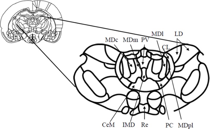
Figure 1.
Schematic diagram (and enlargement) of the medial aspects (Bregma—2.56 mm) of the medial thalamus in the rodent brain. Abbreviations: CeM, center median nucleus, part of the midline nuclei; CL, centrolateral nucleus, part of the intralaminar nuclei; IMD, intermediodorsal nucleus, part of the midline nuclei; LD, laterodorsal nucleus; MDc, central subdivision of mediodorsal thalamus; MDl, lateral subdivision of mediodorsal thalamus; MDm, medial subdivision of mediodorsal thalamus; MDpl, paralamellar subdivision of the mediodorsal thalamus; PC, paracentral nucleus, part of the intralaminar nuclei; PV, paraventricular nucleus, part of the midline nuclei; Re, reuniens. Adapted from Paxinos and Watson (1998).

