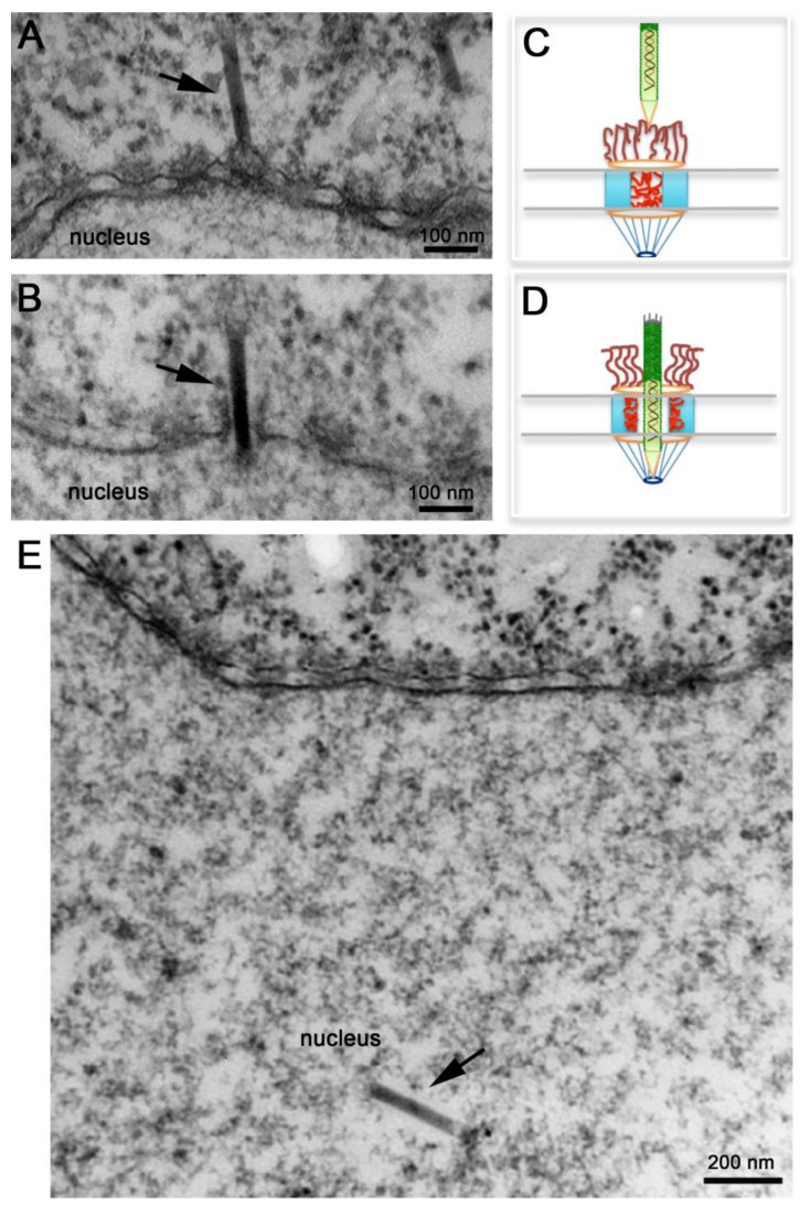
Figure 4.
Nuclear import of AcMNPV Nucleocapsids. Electron micrographs of nuclear envelope cross-sections from Xenopus oocytes that have been microinjected with AcMNPV nucleocapsids and processed for embedding and thin-section electron microscopy. Intact nucleocapsids (arrows) were detected docked at the nuclear pore complex (NPC) prior to nuclear import (A), midway through the NPC (B), and inside the nucleus (E). (C, D) Schematic diagrams of NPCs illustrating “close” and “open” states respectively. In (C) NPC cytoplasmic filaments and nucleoporins within the central channel prevent the passage of the nucleocapsid. In (D) NPC cytoplasmic filaments straighten out, and nucleoporins in the central channel retract towards the body of the NPC to free up the passageway for the nucleocapsid to cross the NPC.

