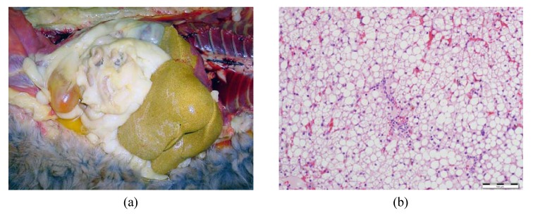
Figure 4.
The liver of an obese cat with hepatic lipidosis. (a) In situ liver of an obese cat with hepatic lipidosis; note the yellow discoloration and hepatomegaly. (b) Liver tissue of an obese cat with hepatic lipidosis, showing diffuse cytoplasmic vacuolization of the hepatocytes representing fat accumulation (H & E staining, original magnification: 20×).

