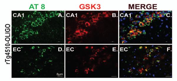
Figure 12. Phosphorylated tau co-localized with activated GSK3 in rTg4510 mouse brain.

Tau at pS202/T 205 (AT8; green) is co-localized with phosphorylated GSK3 α/β (red) in the neuronal cell bodies of CA1 (A-C) and entorhinal cortex (D-F) in rTg4510 mice infused with OLIGO. DAPI was used to stain nuclei (blue). The scale bar represents 8 μm for all panels.
