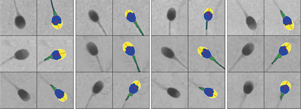
Figure 1.

Paired original (left panel) and false-colour-coded digitized (right panel) images, showing the sperm midpiece (green with axis) and head subdivided into acrosomal area (yellow) and post-acrosomal region (blue). The major (length) and minor (width) axes are the red lines and perimeter is in white.
